The AutoDrive Challenge™ donation Chevy Bolt EV’s are in final stages of inspection and completion prior to being shipped to you!
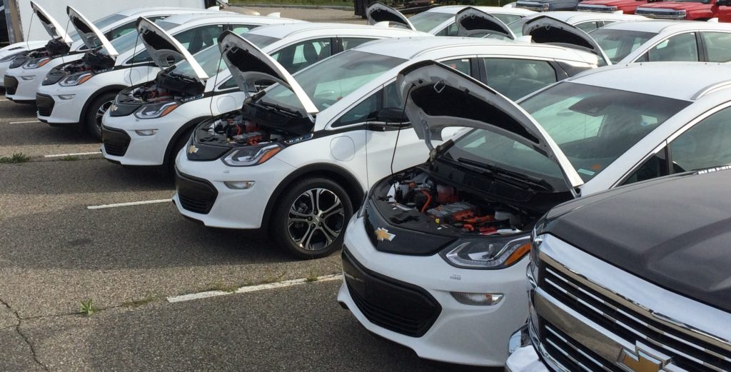


The AutoDrive Challenge™ donation Chevy Bolt EV’s are in final stages of inspection and completion prior to being shipped to you!

Kettering University is proud to be one of eight schools nationwide to be selected to participate in the Autodrive Challenge.
Kettering University is one of eight universities around the world selected to participate in the Society of Automotive Engineers’ (SAE) AutoDrive™ Challenge – an international autonomous vehicle competition.
This newly established, three-year competition will task faculty and students at some of the world’s top universities with developing and demonstrating a fully operational autonomous driving passenger vehicle. The technical goal of the competition is to navigate an urban driving course in an automated driving mode by year three.
 https://news.kettering.edu/news/kettering-university-selected-participate-international-autonomous-vehicle-challenge
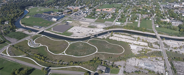
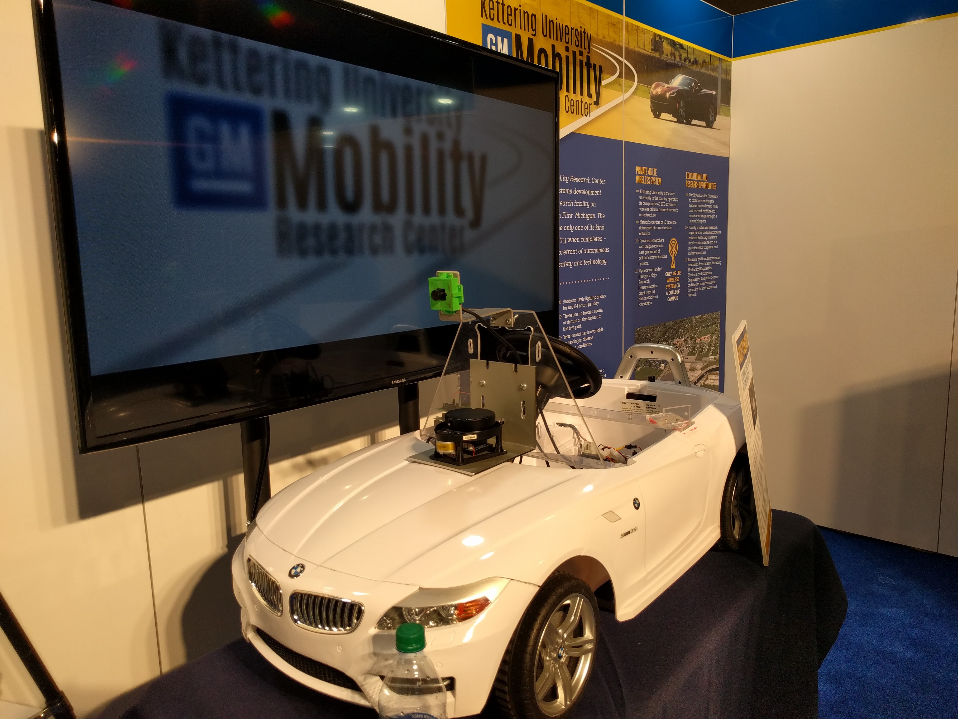
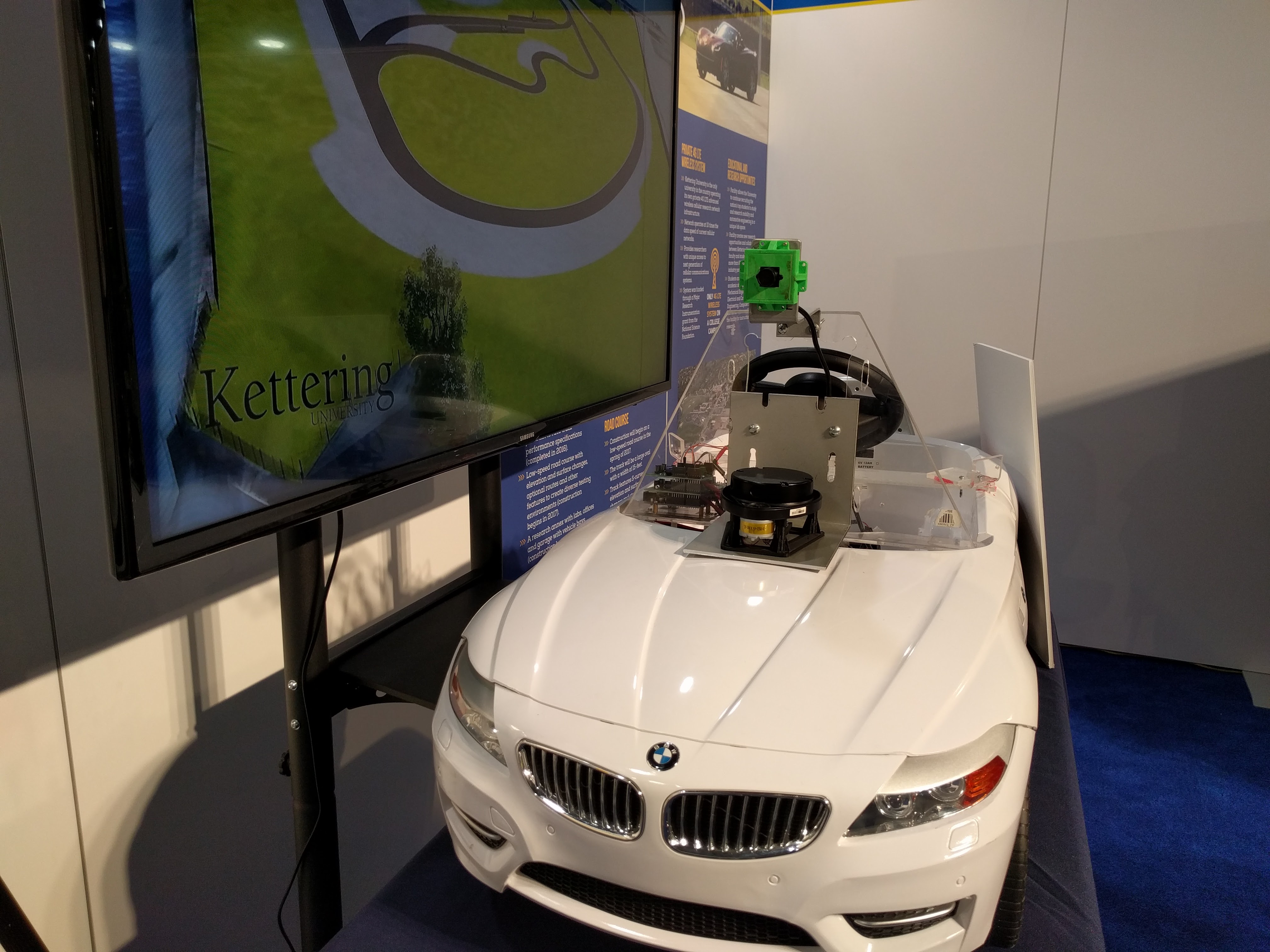
Kettering is the only university in the country with an industry-standard proving ground physically located on its campus. Phase one of the Kettering University GM Mobility Research Center (MRC) was completed in the fall of 2016 and phase two is scheduled to be completed in 2017.
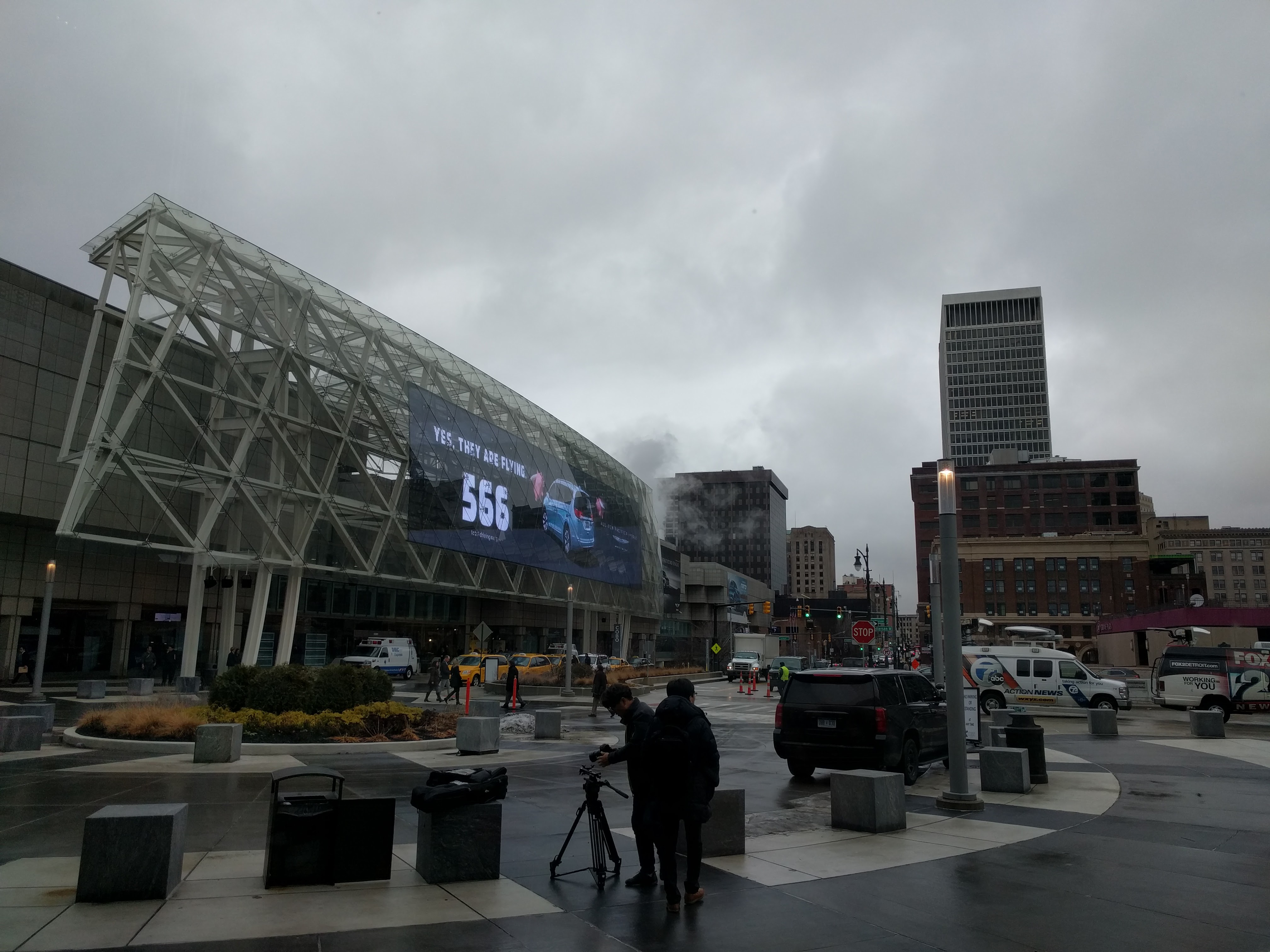
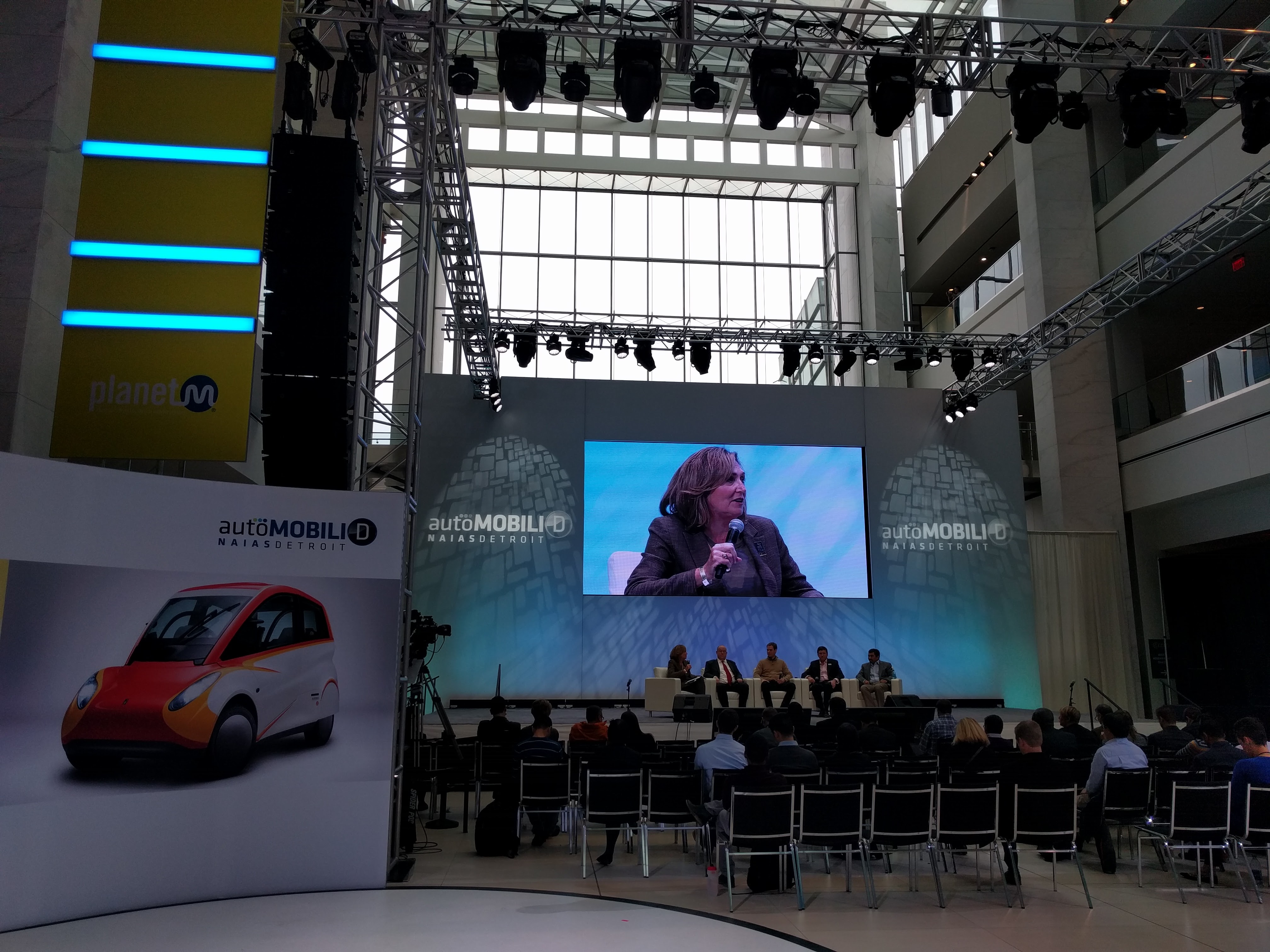
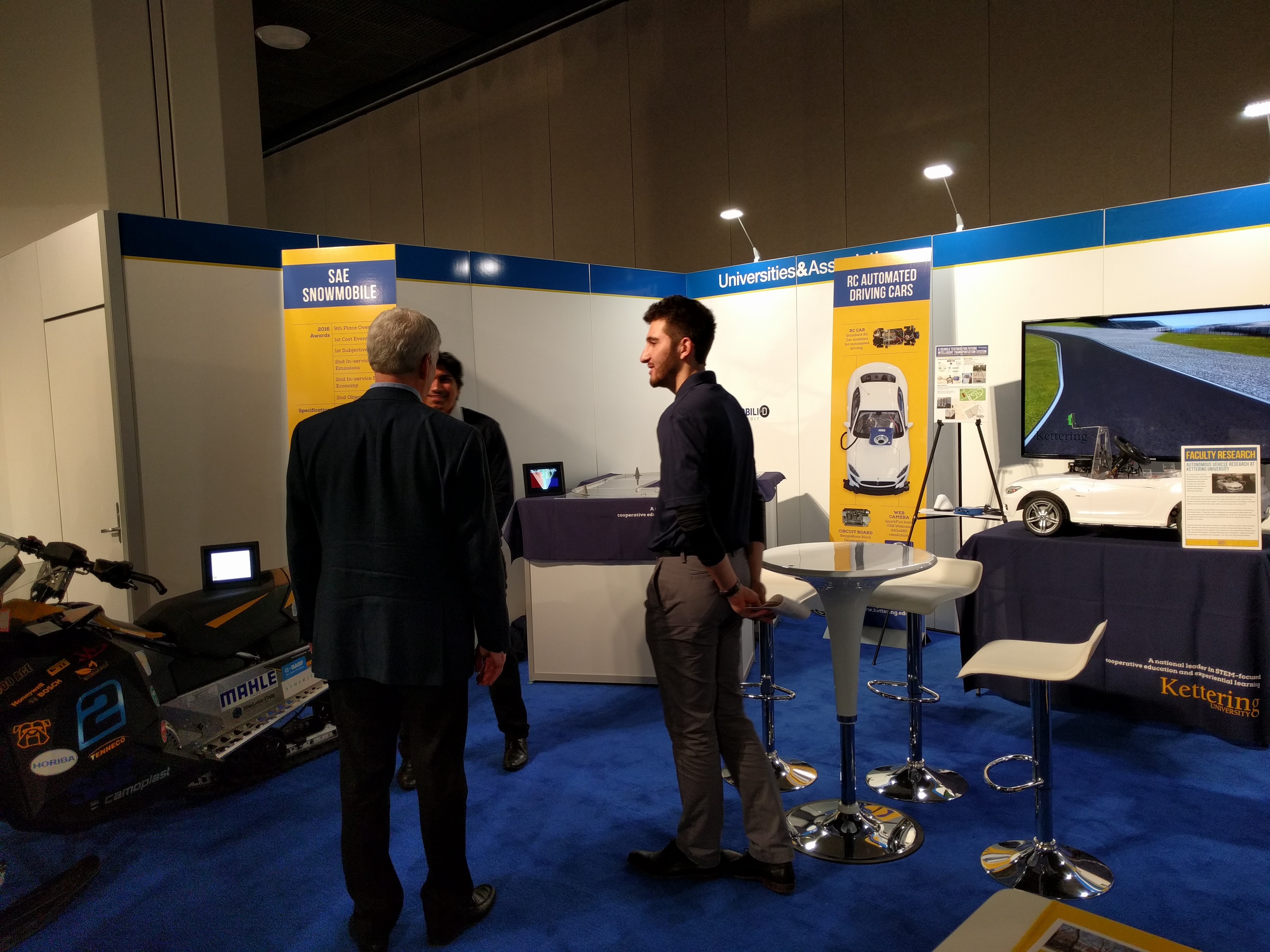
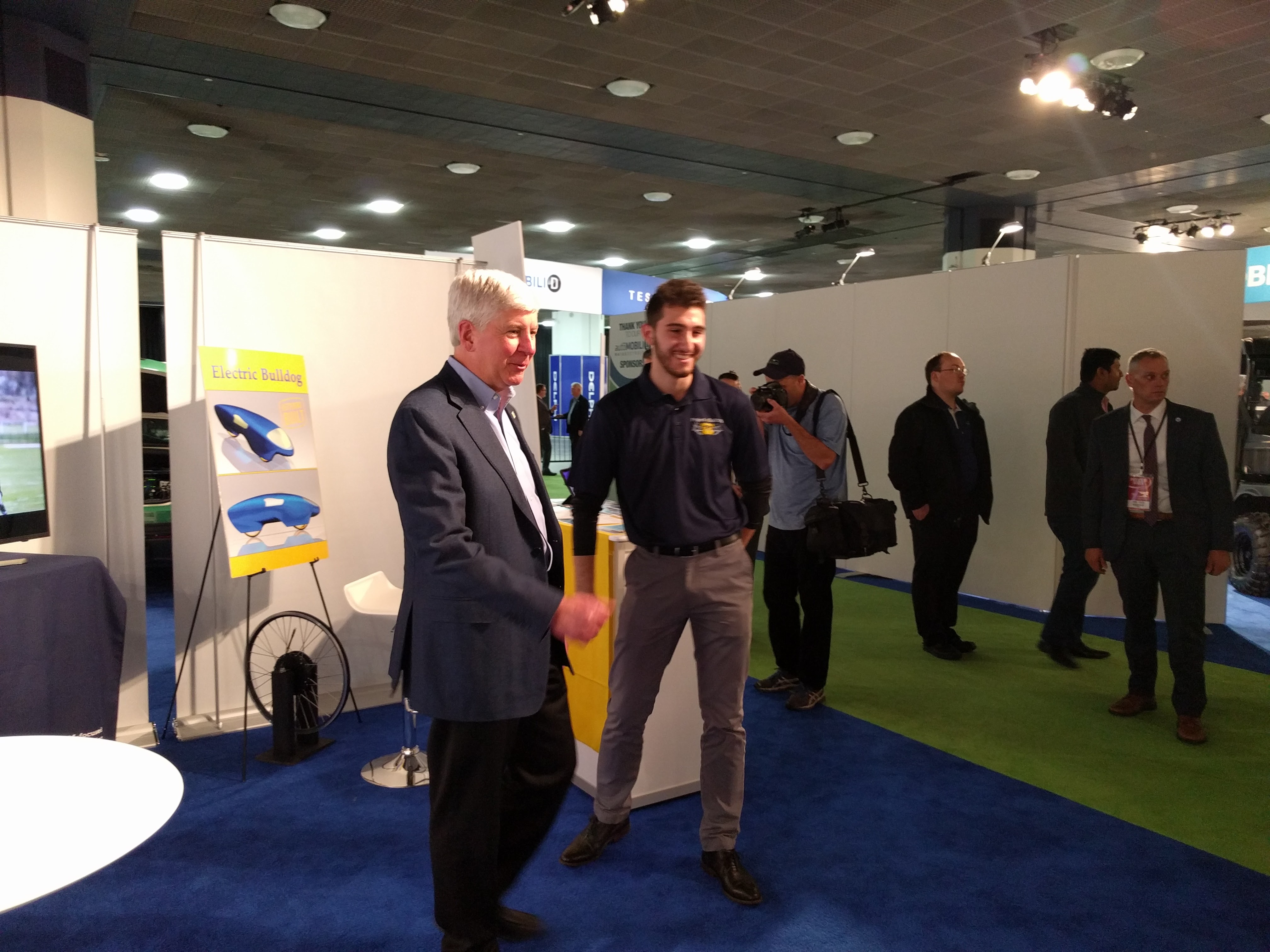
Research on Autonomous Driving from MIR Lab was presented in AutoMobili-D at Cobo Center Atrium, Detroit as part of Detroit Auto Show 2017.
My project among others funded by an NSF grant was introduced on the Flint Journal, a local newspaper. It is not 100% accurate descriptions about me and the project. One example is that it was not 10 years ago when I studied at Texas A&M University.
Kettering University receives half a million dollars in National Science Foundation grants
This one is on the school news coverage. Again the title is not. It is NOT “MRI” research. The grant name is “MRI” which stands for Major Research Instrument.
NSF grant will support high resolution MRI research
ABC12 news
Kettering University receives grants from National Science Foundation
Computer Engineering Summer Day Camp is one of pre-college programs that GM Foundation decides to fund. The summer camp started from 2011 and has been successful.
I am honored to have this great opportunity thanks to many people including my graduate advisor and his team. This would not be possible without full support of our department head and the office of sponsored office at Kettering.
MRI: Development of High-Throughput ad High-Resolution Three-Dimensional Tissue Scanner with Internet-Connected 3D Virtual Microscope for Large-Scale Automated Histology
The objective of this program is to develop a high-throughput and high-resolution 3D tissue scanner with an Internet-connected 3D virtual microscope for large-scale automated histology. The instrument will digitize a wide variety of organs and generate 3D data sets. A data processing pipeline will automatically convert the raw data sets into aligned volumetric data sets without human intervention. Therefore immediate access to the large-scale histological data will be possible for a wide variety of research and research training communities.
The intellectual merit is as follows. Large-scale output data of the instrument will be easily acquired and shared through the fast scanner, data processing pipelines, and web-based visualization framework. In the past, investigation of biological organs were limited to either gross system-level studies or highly detailed microscopic analysis of small subsets of the organ. The new tissue scanner will digitally restore tissue structure in 3D with a specialized visualization framework that helps researchers to fully understand function of biological organs.
The broader impacts are three folds: (1) Obtaining digitized high-resolution data for multi-scale investigation of biological organs at a high acquisition rate will open up new areas of research in the community. (2) For education infrastructure, microscopic atlases of whole biological organs can serve as educational materials for students and educators at all levels. (3) For dissemination, the project results will provide research communities with a new 3D tissue scanner with a virtual microscope by which researchers can obtain digitized 3D tissue data in sub-micron level resolution from a tissue sample.
http://www.nsf.gov/awardsearch/showAward?AWD_ID=1337983
During Homecoming Weekend, posters from the M:IR lab were presented at the Celebrate Achievement Expo from 9-11 a.m. May 18.
Microsoft kinetic sensor for real-time color tracking robot
Matthew Clark, David Feldpausch, and Girma Tewolde, Department of Electrical and Computer Engineering
Low-cost dashboard for an electric scooter
Trifon Tsekov, Girma Tewolde, Jae Kwon and Kevin Bai, Department of Electrical & Computer Engineering
Development of a modular LED display controller
Dan Gudorf and Girma Tewolde, Department of Electrical & Computer Engineering
Kettering University Robotics Team – Intelligent ground vehicle competition
Jorge Horcasitas, Lex Lombardi and Girma Tewolde, Department of Electrical & Computer Engineering
Design and implementation of LED dimming system with intelligent sensor module
Jaerock Kwon, Department of Electrical and Computer Engineering
 |
| Trifon, Reza(Haptics Lab member), Girma, Jae, Jongil (Joe), Seokju (Sean). |
The Mobile: Intelligent Robotics (M:IR) Lab has a new space. At the same time, two new graduate students, Seokju Lee and Jongil Lim who came from Korea, joined the M:IR Lab. Welcome to the Mobile Intelligent Robotics Lab and Kettering University.
To celebrate the new space we had a small kick-off meeting with other faculty members. Thank you all!
 |
| Jim is giving us a short speech to welcome the new grads. |
 |
| Kevin is asking if the bread is made by Girma’s wife. |
 |
| Mo also joined to celebrate this new space with his daughter. |


Next week, the ECE department hosts Computer Engineering Day Camp for high school students. The program has two different sections: 1) Mobile robot, 2) Smart phone programming. But in the last day, two sections will be integrated into one to control Lego Mindstorm Robots with Smart devices through the Bluetooth technology.
I am responsible for the second section, smart phone programming. To be precise, smart device programming is better term since I am going to introduce some additional tablet devices powered by Android.
App Inventor for Android will be used for programming The block based graphical programming tool, similar to Scratch, was originally from Google Labs. Now it is maintained by MIT.
For more details such as lecture materials, you can visit here.
The Journal of Visualized Experiments (JoVE) is a peer reviewed, PubMed-indexed video journal. My research on 3D tissue scanner was publicshed on the journal. Specimen Preparation, Imaging, and Analysis Protocols for Knife-edge Scanning Microscopy. Here is an abstract of it.
Major advances in high-throughput, high-resolution, 3D microscopy techniques have enabled the acquisition of large volumes of neuroanatomical data at submicrometer resolution. One of the first such instruments producing whole-brain-scale data is the Knife-Edge Scanning Microscope (KESM)7, 5, 9, developed and hosted in the authors’ lab. KESM has been used to section and image whole mouse brains at submicrometer resolution, revealing the intricate details of the neuronal networks (Golgi)1, 4, 8, vascular networks (India ink)1, 4, and cell body distribution (Nissl)3. The use of KESM is not restricted to the mouse nor the brain. We have successfully imaged the octopus brain6, mouse lung, and rat brain. We are currently working on whole zebra fish embryos. Data like these can greatly contribute to connectomics research10; to microcirculation and hemodynamic research; and to stereology research by providing an exact ground-truth.
In this article, we will describe the pipeline, including specimen preparation (fixing, staining, and embedding), KESM configuration and setup, sectioning and imaging with the KESM, image processing, data preparation, and data visualization and analysis. The emphasis will be on specimen preparation and visualization/analysis of obtained KESM data. We expect the detailed protocol presented in this article to help broaden the access to KESM and increase its utilization.